Education
Integrating Augmented Reality in vascular neuroanatomy teaching for medical students.
Integrating Augmented Reality in Vascular Neuroanatomy Teaching for Medical Students

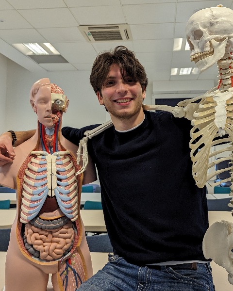
Ally Fattal, Third year medical student at King's College London
Presenting author
King's College London
Presenting Author(s)
Introduction: Understanding vascular neuroanatomy is crucial for medical students, as it plays a vital role in diagnosing and managing neurological and neurosurgical conditions. This study investigates the effectiveness of augmented reality (AR) in enhancing the comprehension of vascular neuroanatomy and spatial vascular anatomy in a cohort of medical students.
Methods: A total of 100 first-year medical students at Imperial College London were randomly assigned to two groups: one utilizing simXAR AR technology for learning vascular neuroanatomy and the other receiving traditional didactic instruction. The AR group engaged with 3D interactive models that allowed them to visualize vascular structures and their spatial relationships dynamically. Both groups completed pre- and post-assessments that included objective tests and a spatial orientation questionnaire. Statistical analyses were performed using paired t-tests to evaluate differences in understanding and spatial awareness.
Results: The results indicated that the AR group exhibited a significant improvement in understanding vascular neuroanatomy. Pre-test scores for the AR group averaged 68% (SD = 12.3), while post-test scores increased to 88% (SD = 9.4), demonstrating a 20% improvement (p < 0.001). Conversely, the traditional group showed minimal improvement from 67% (SD = 11.5) to 71% (SD = 10.8) (p = 0.09). Additionally, the AR group reported enhanced spatial comprehension, with scores increasing from 3.4 (SD = 0.8) to 4.6 (SD = 0.6) (p < 0.001).
Conclusion : The incorporation of augmented reality in teaching vascular neuroanatomy significantly enhances student understanding and spatial awareness. These findings support the integration of AR technology into medical education to improve learning outcomes in complex anatomical subjects.

.jpg)